Unblocking in Here Be Dust
- March 20, 2016, 11:25 a.m.
- |
- Public
I had my follow-up ultrasound on March 18 for my port-related blood clot. The upshot: My subclavian vein is still partially blocked, but it is well on its way to being fully open again.
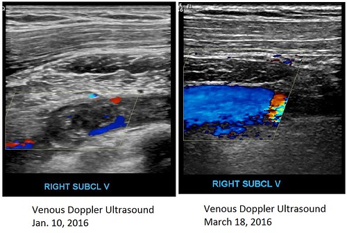
From my understanding of the video that I posted in this entry (which, like most videos about DVTs (blood clots), deals with the leg, but basic concepts remain the same), the colored patches indicate blood flow. (The explanation of color begins at around the 13:10 mark.) Red means inflow; blue means outflow. As shown in my Jan. 10 ultrasound, most of that flow had been blocked by the clot. By the time of Friday’s follow-up ultrasound, much of it had been restored.
Or, as my ultrasound report put it, “Partial recanalization of the chronic deep venous thrombosis within the right axillary/subclavian vein junction.” I’m not out of the woods yet, but I’m getting there.
The ultrasound tech remarked on my small veins when she tried to visualize them, which confirms my statements that they’re persnickety. She had particular trouble near my shoulder, I think with my cephalic vein.
I estimate that my venous swelling has decreased by about 95 percent. These days I’m wearing the Koi design LympheDIVAs compression sleeve. My GP’s initial response had been alarm (“What did you do to your arm?! You got tattooed?!”) – she had interrupted herself while examining my partner, two days before my own appointment. It was the first time she had seen me since she had stopped by my hospital bed in January. The sleeve has gotten me compliments from people, including other health care professionals, on my “tattoo.”
Fortunately, the swelling is down enough so that my right arm can again be used for blood pressure. On the day of my appointment it was 112/72. I’ll take it. My GP said that I can also have blood draws taken from that arm again. I’m hoping for better results on that front next month, when I’m due for blood work at my oncologist’s office. Lymphedema risk put my left arm out of commission for needles (of course that would be the arm with the easier veins), and we had conceded failure after four attempts on my right hand (and my almost passing out) back in January.
I had some wait time before I could register and then before I could be seen for the ultrasound, but this time I had my new tablet with me. I don’t have a smartphone; friends had posted updates for me on Facebook during my hospital stay. I was and am very thankful for their help back then, but I felt it was time for me to have better online access. As a caregiver I’ve brought my laptop and used the hospital’s WiFi to communicate, but I wanted something that I could always keep with me as a matter of course – without investing in a smartphone and the contract that goes with it.
My other main reason for getting a tablet was so that I could play with ArtRage on the go. :-)
I took advantage of a sale and picked up this, along with a stylus, microfiber cloth, micro-SD card, and protective case. Unless I want to get online, I have the tablet’s WiFi turned off to conserve battery strength.
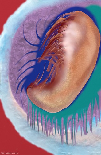
I’m still learning the Android version of ArtRage, which is not as extensive as the desktop version. It still has plenty to offer, especially for a doodler like me. While waiting at the hospital I came up with this little abstract; I added the text at home using my laptop:
The tablet has a 5-megapixel camera on the back and a 2-megapixel camera on the front. I used the front-facing camera to take this shot:

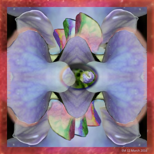
Our yard has now exploded with snapdragons (Linaria canadensis, a.k.a. toadflax). I used my Plugable digital USB microscope to take this shot, then played with it and others using the full-featured ArtRage:
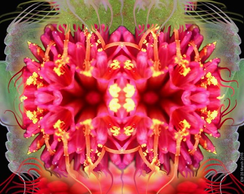
To celebrate Spring I used images (also captured with the microscope) of Emilia fosbergii (a.k.a. Florida Tasselflower and Cupid’s Shaving Brush) from our back yard, and a new leaf from our dwarf elm tree. Having occurred at 4:30 a.m. UTC (12:30 a.m. Eastern time), this marks the earliest spring since 1896.
The Citizen Science Trailblazer 2 Project from Cancer Research UK is now live. Participants (of which I am one) analyze images to find cancer cells after completing a tutorial. Says the website, “With your help, we’ll take one step further towards creating a brand new method for analysing cancer data on a huge scale.”



Loading comments...